
Grant Smith, Jocelyn Williams, Helen Haines
Abstract
Located in northern Belize, the ancient Maya site of Ka’kabish had a long occupational history spanning the Middle Formative period (800-600 BC) into the Postclassic period (900-1500 AD). Excavations at Ka’kabish revealed eight plazas and courtyards, the largest plaza being Group D. To the south of Group D were several chultuns, or subterranean chambers, which functioned as burial sites for the occupants of Ka’kabish; all chultuns contained multiple interments. For my master's thesis, I was tasked with examining the material from the chultuns, identifying MNI and investigating diet and health using both stable isotope and osteological analyses. As is typical of skeletal material in the Maya area, preservation of the human remains from the chultuns is poor and the burials are commingled. Based on the osteological examination, I determined an MNI of 28 individuals—18 adults, 5 subadults and 5 neonate/perinates. The poor preservation and fragmentary nature of the commingled assemblage made the identification of sex and pathological conditions difficult and impossible for most of the skeletal material. In one chultun, however, the preservation was very good and it was possible to observe pathological changes on numerous skeletal elements corresponding to at least two adult individuals. Lesions consistent with osteoarthritis, infection, metabolic disease and congenital defects are present. The pathological conditions are quite variable and do not clearly correspond to a systemic disease or a single etiology. The purpose of this poster is to illustrate the lesions, present differential diagnoses, and discuss possible impacts of these diseases.
References Cited
- Appleby, J., Thomas, R., & Buikstra, J. (2015). Increasing confidence in paleopathological diagnosis–Application of the Istanbul terminological framework. International Journal of Paleopathology, 8, 19-21.
- Aufderheide, A., Rodriguez-Martin, C. (1999). The Cambridge Encyclopedia of Human Paleopathology. Cambridge University Press.
- Dounis, E. (1978) Sacrococcygeal Agenesis: A report of four new cases, Acta Orthopaedica Scandinavica, 49, 475-480.
- Duncan, P. A., Shapiro, L. R., & Klein, R. M. (1991). Sacrococcygeal dysgenesis association. American Journal of Medical Genetics, 41(2), 153-161.
- Haines, H.R., Sagebiel, K.L., & Belanger, C. (2017). Is and Isn’t produce each other: An Unusual Architectural Amalgamation at Ka’kabish. Research Reports in Belizean Archaeology,14, 123-134.
- McLellan, A., Haines, H.R. (2013) Casting a Light in the Wilderness: The Ancient Maya site of Ka’Kabish, in Northern Belize. Research Reports in Belizean Archaeology,10,189-200.
- Ortner, D. J. (2003). Identification of Pathological Conditions in Human Skeletal Remains. Academic Press.
Images (clicking on image will take you to large version of file)
.jpg)
Figure 1. Adult Sacrum: Shape abnormality, absence of S3-S5 (dorsal view), compared to normal sacrum
.jpg)
Fig 2. Adult Sacrum: Shape abnormality, absence of S3-S5, ventral view
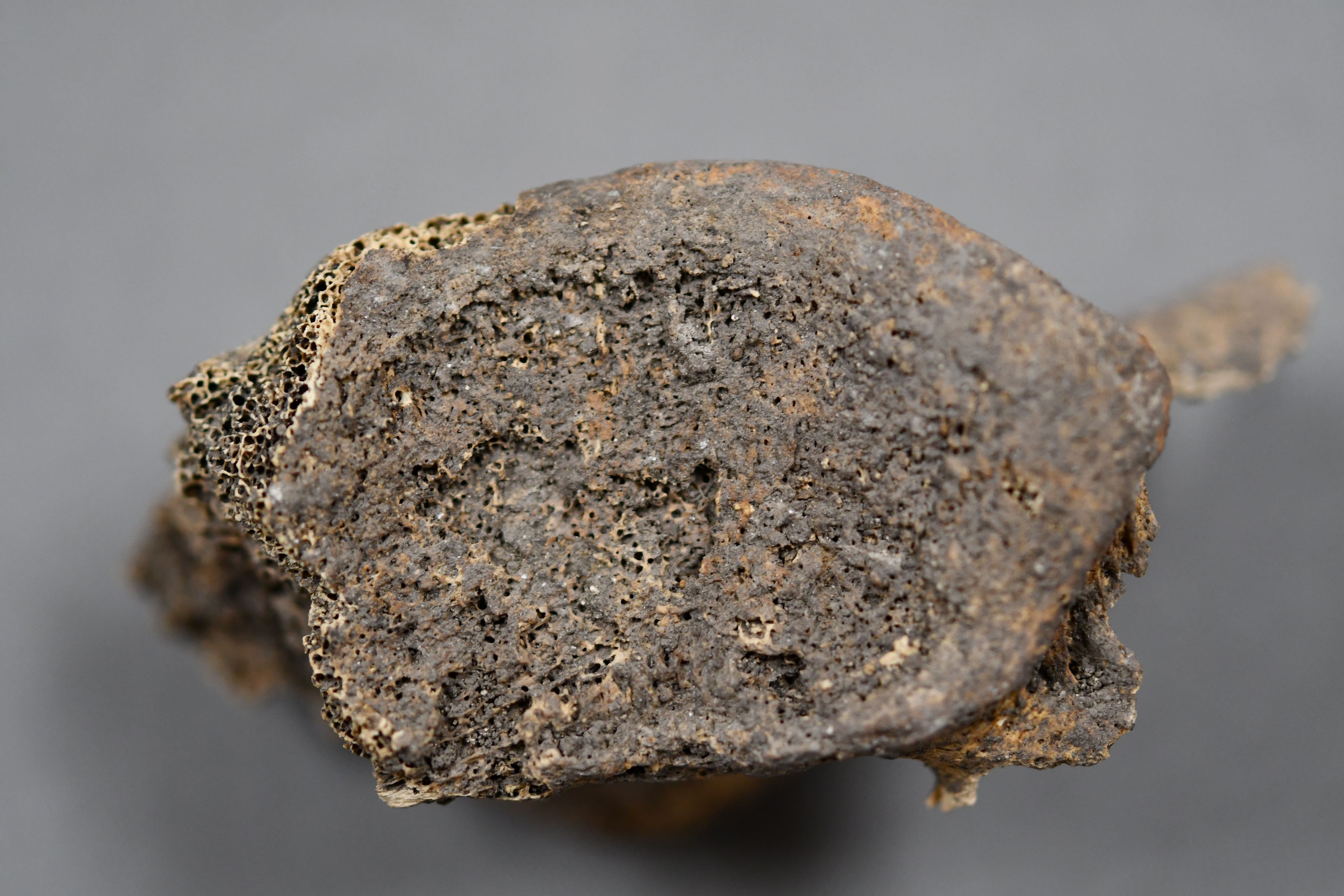
Fig 3. Adult Sacrum: Shape abnormality, superior view
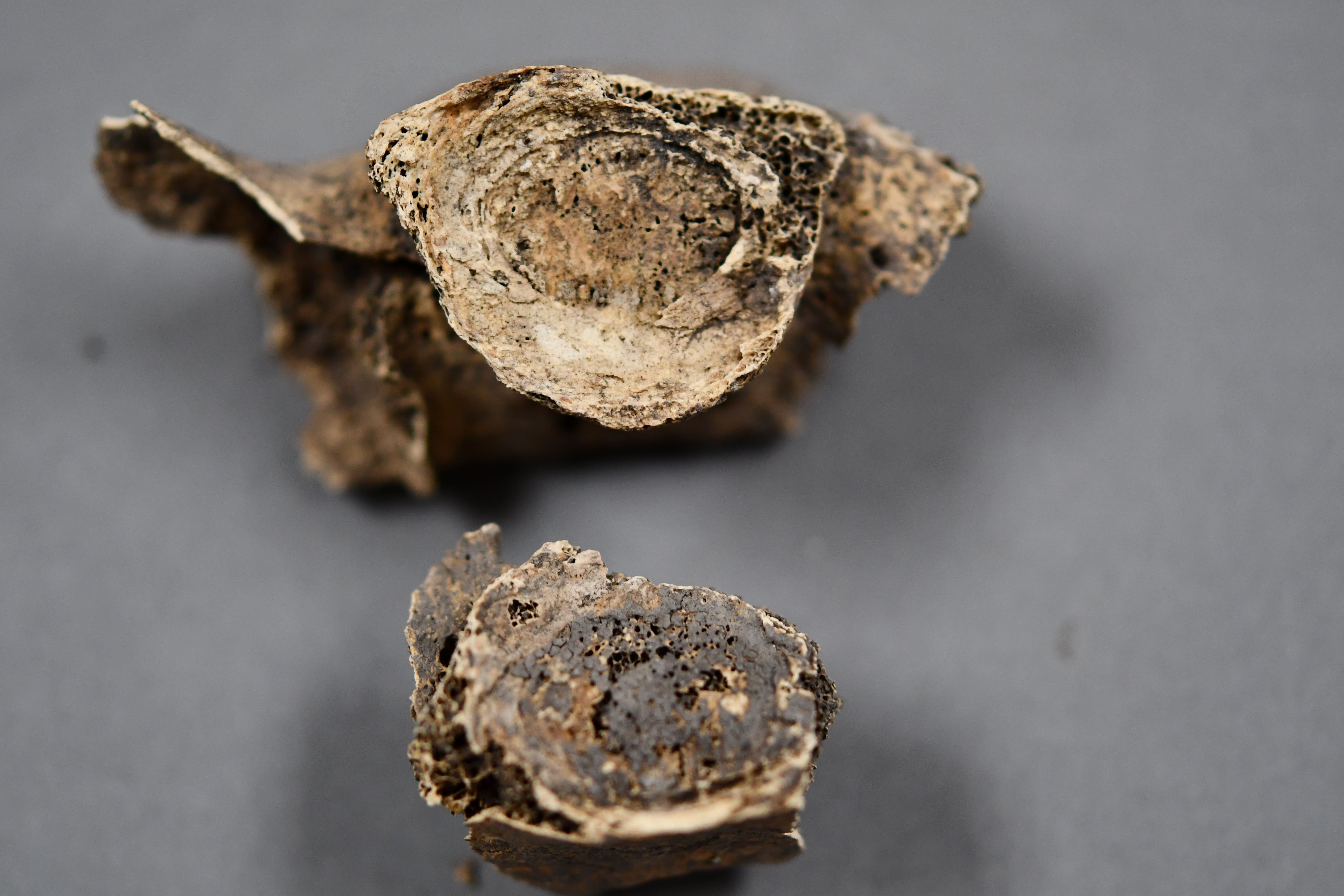
Fig 4. Adult Sacrum: Shape abnormality, absence of S3-S5, inferior view
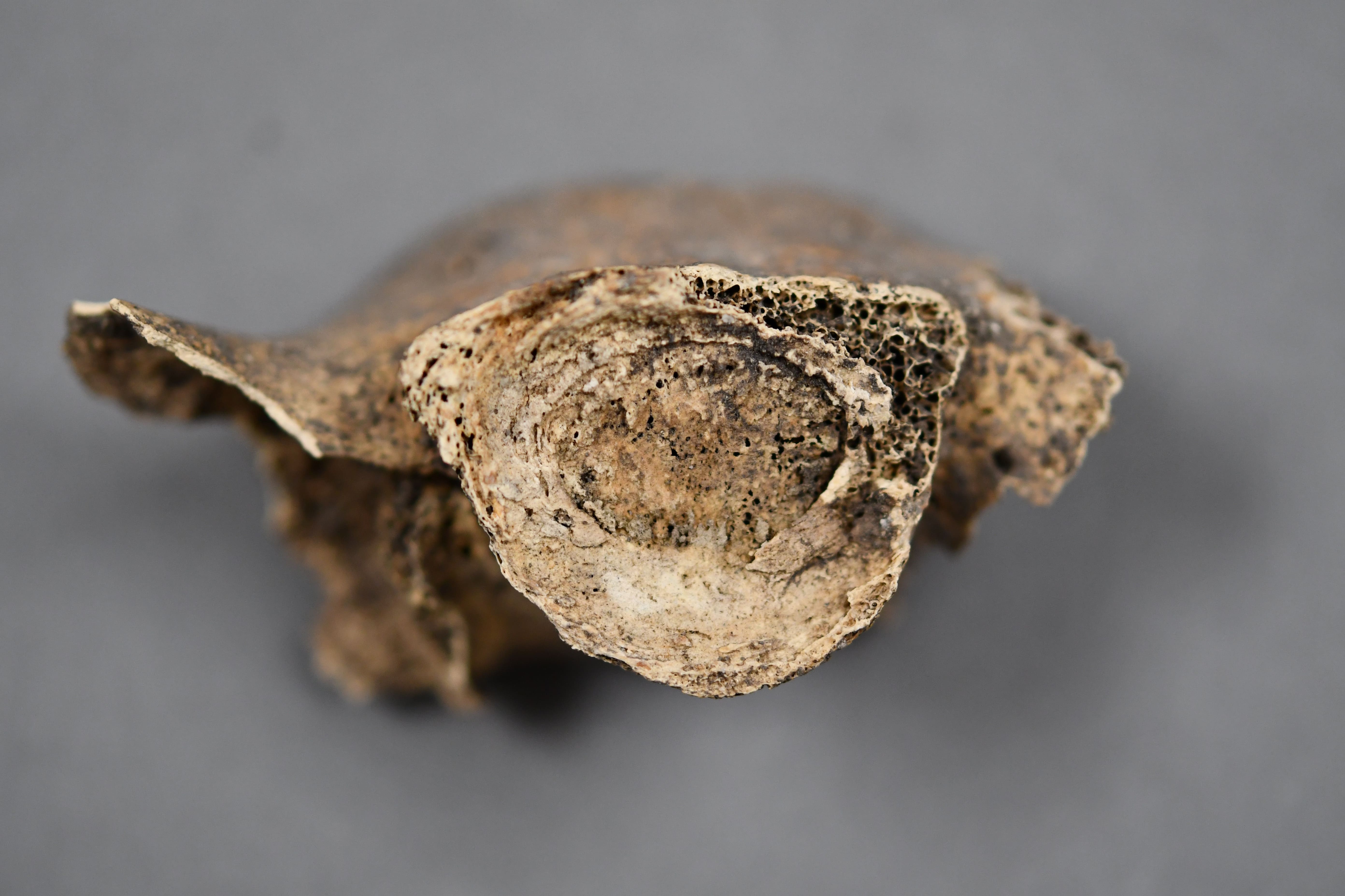
Fig 5. Adult Sacrum: Shape abnormality, S1 inferior view
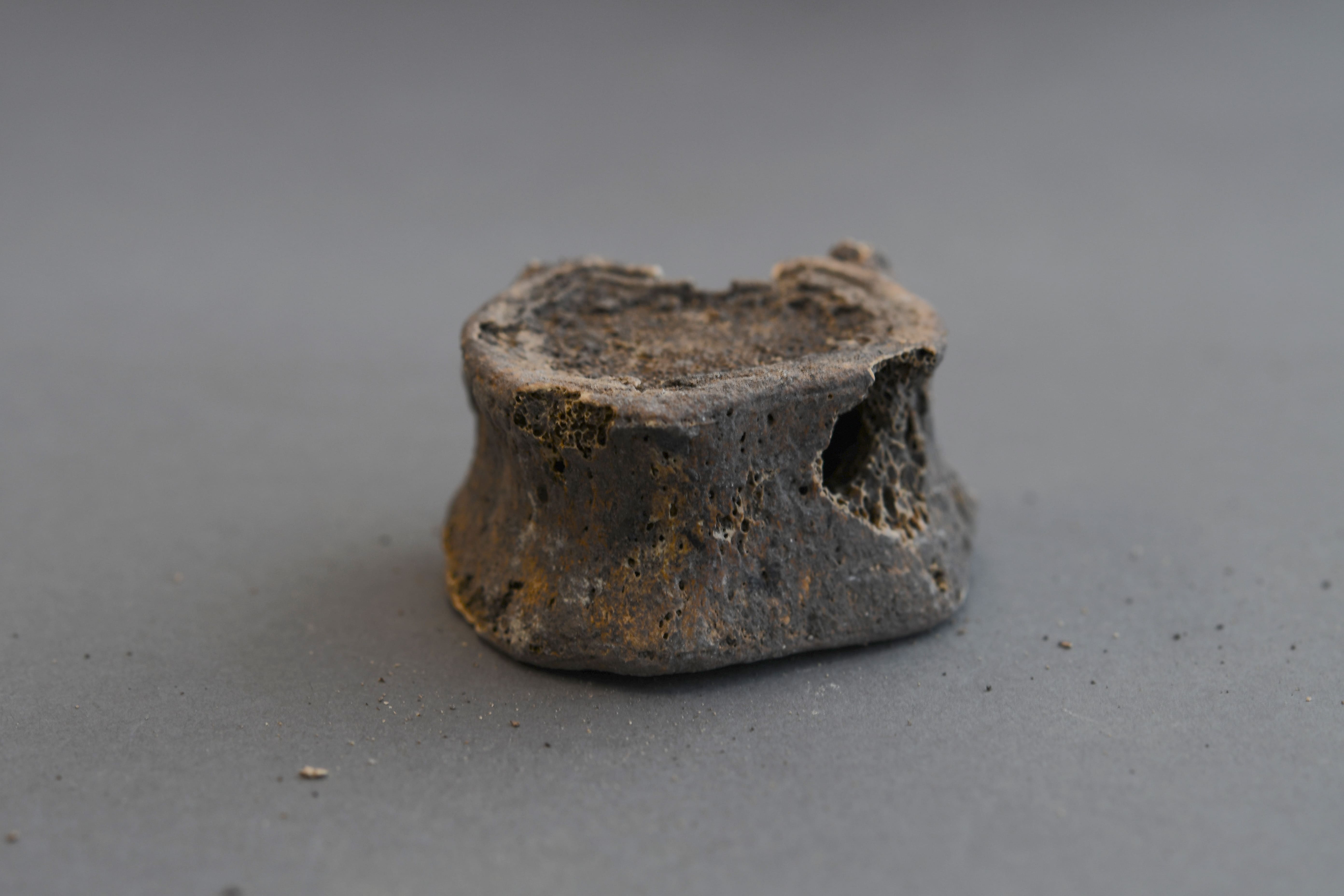
Fig 6.T11 Anterior
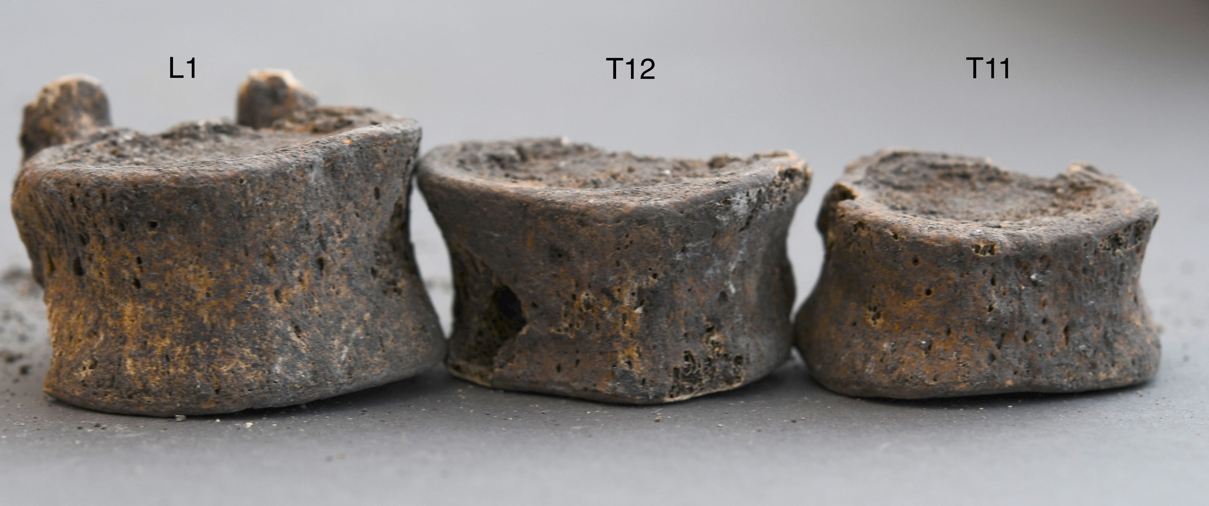
Fig 6. T11, T12, L1 Anterior
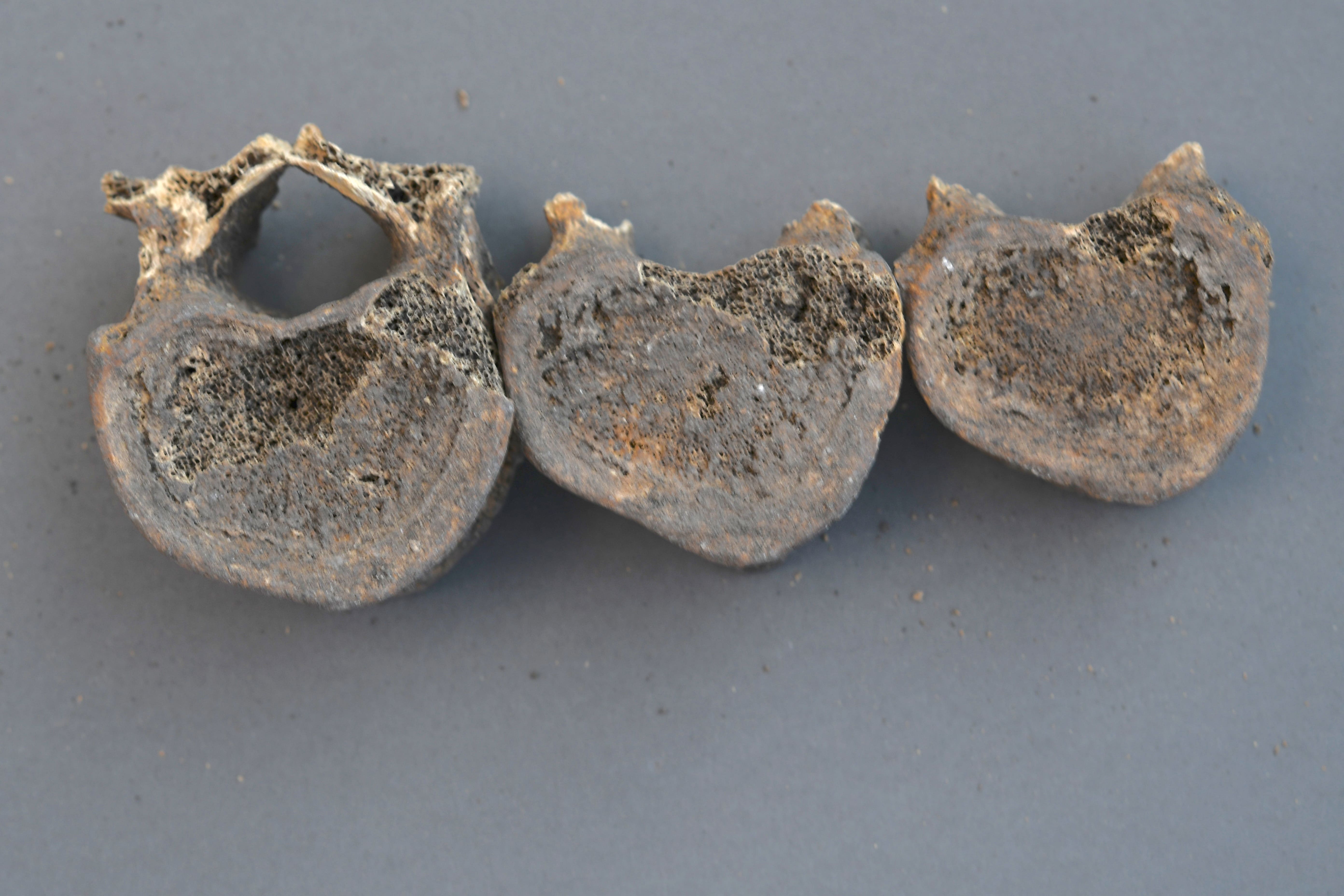
Fig 6. T11, T12, L1 Superior
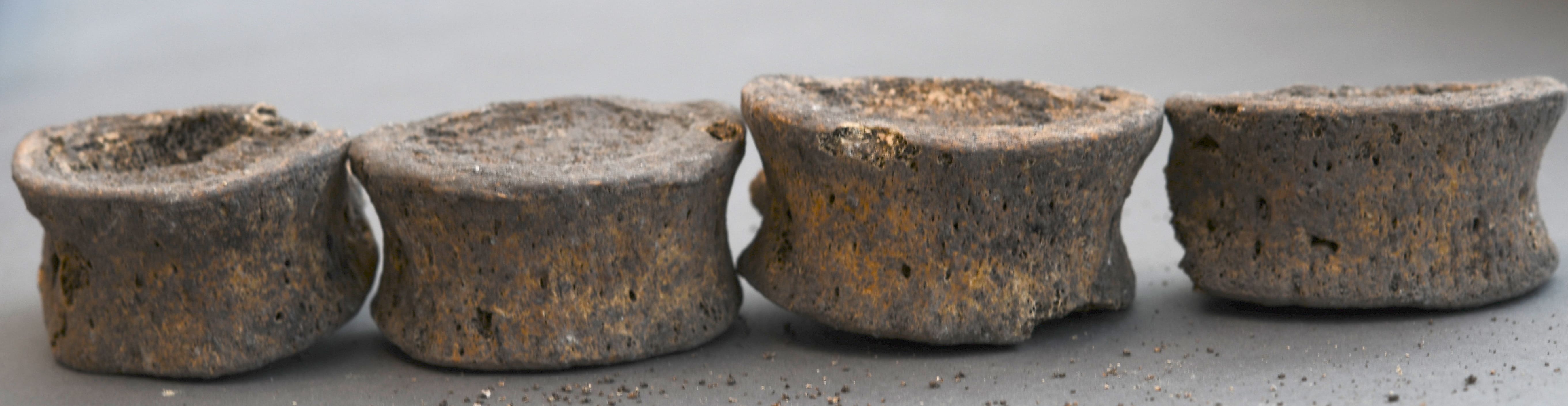
Fig 6. T11, T12, L1, L2 Anterior
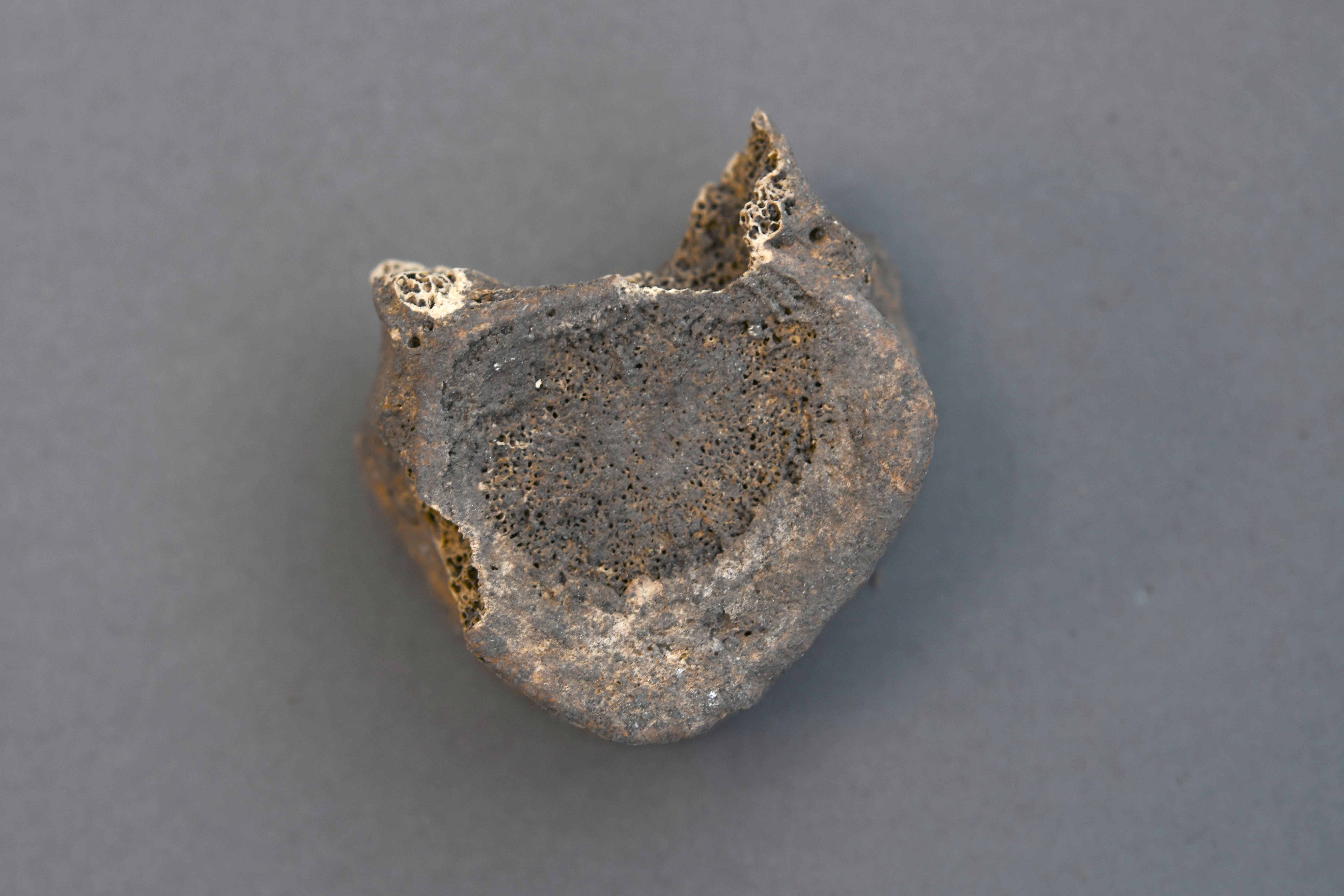
Fig 6. T12 Inferior
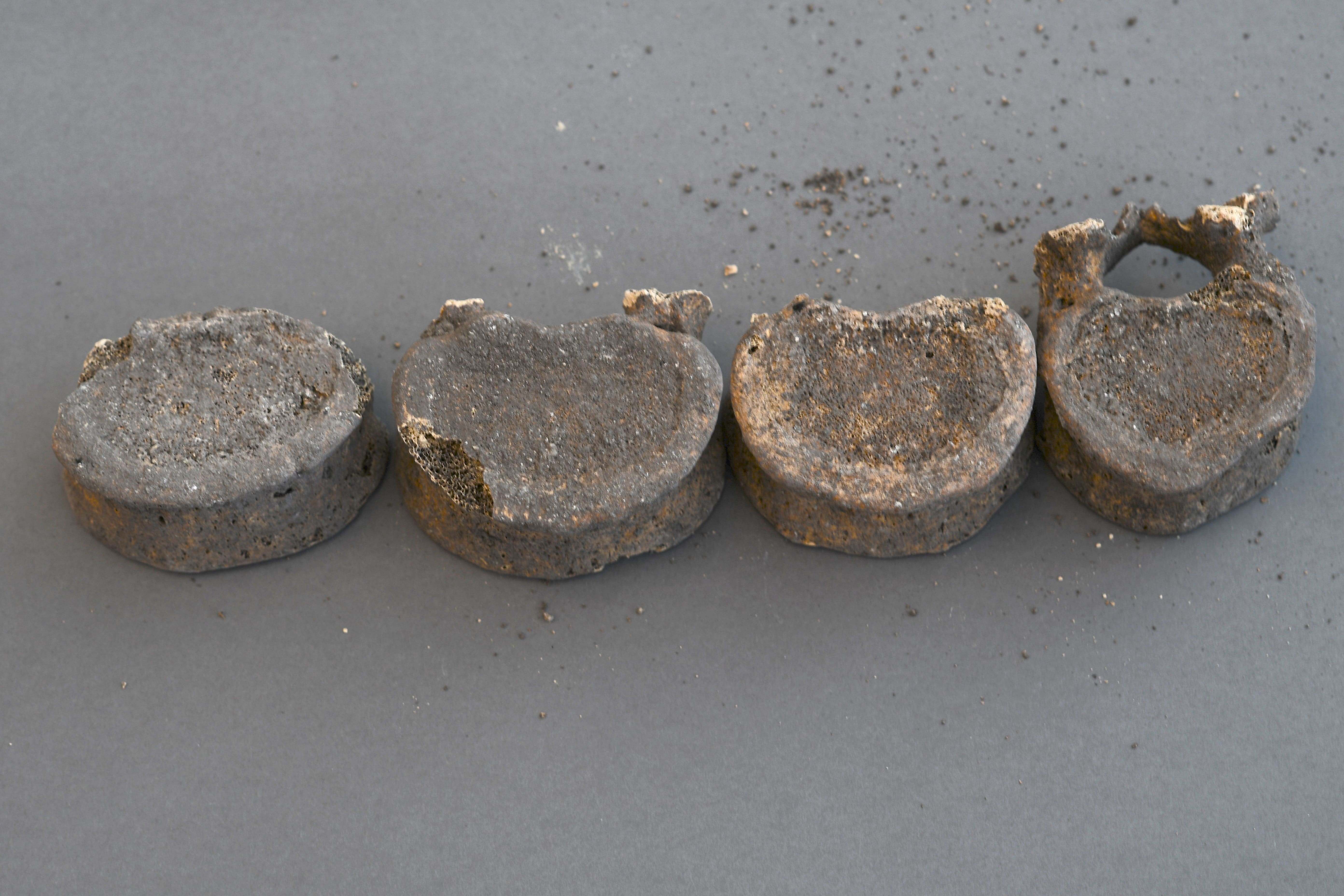
Fig 6. T12, L1, L2, L3, L5 Inferior
.jpg)
Fig 6. Thoracic Comparison
.jpg)
Fig 6. Thoracic Superior
.jpg)
Fig 6. Thoracic Inferior
%20(1).jpg)
Fig 6. Thoracic Vertebrae (T11)
%20(1).jpg)
Fig 6. Thoracic Vertebrae (T12)
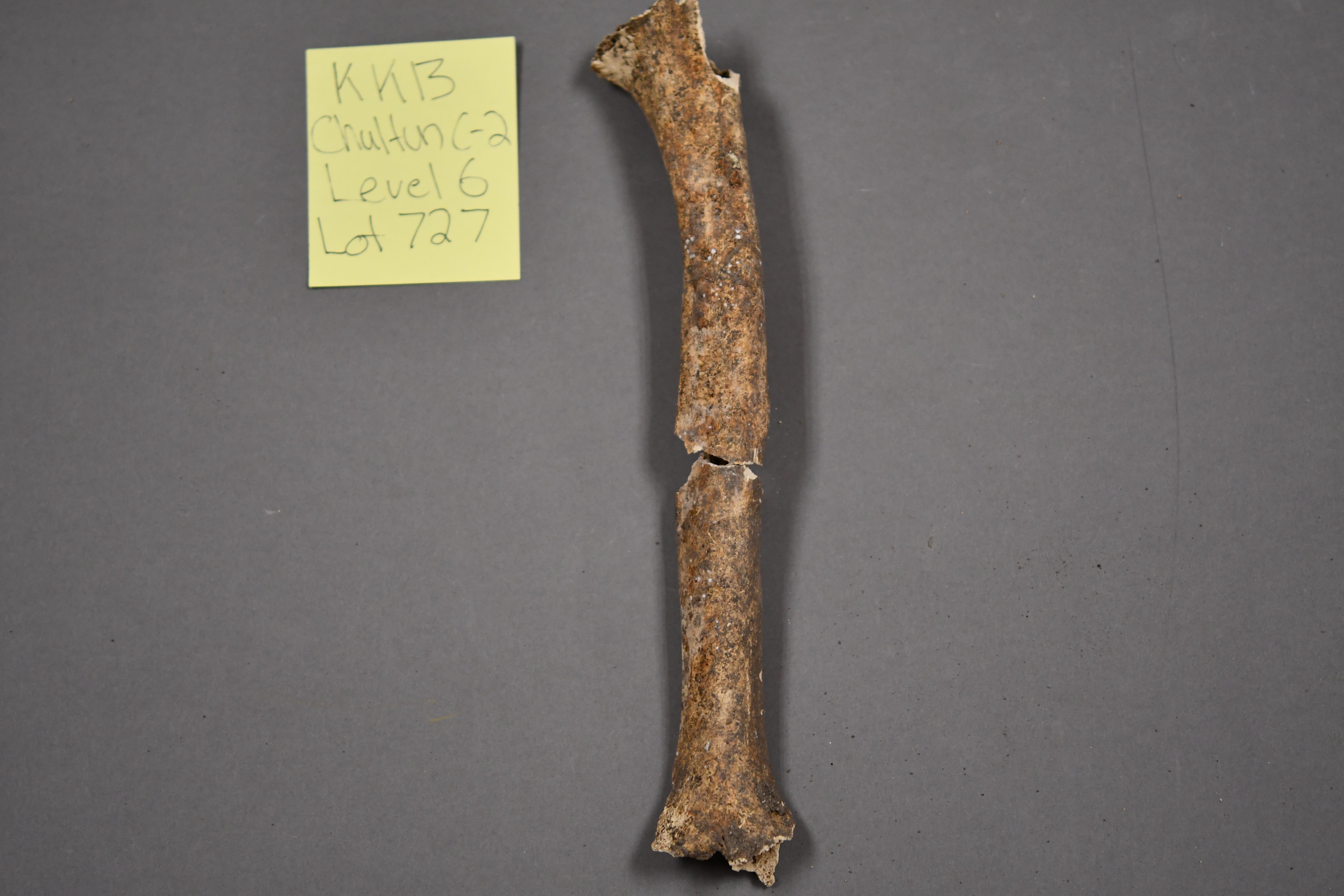
Fig 7. Adult Left Humeri: Bowed/Swollen
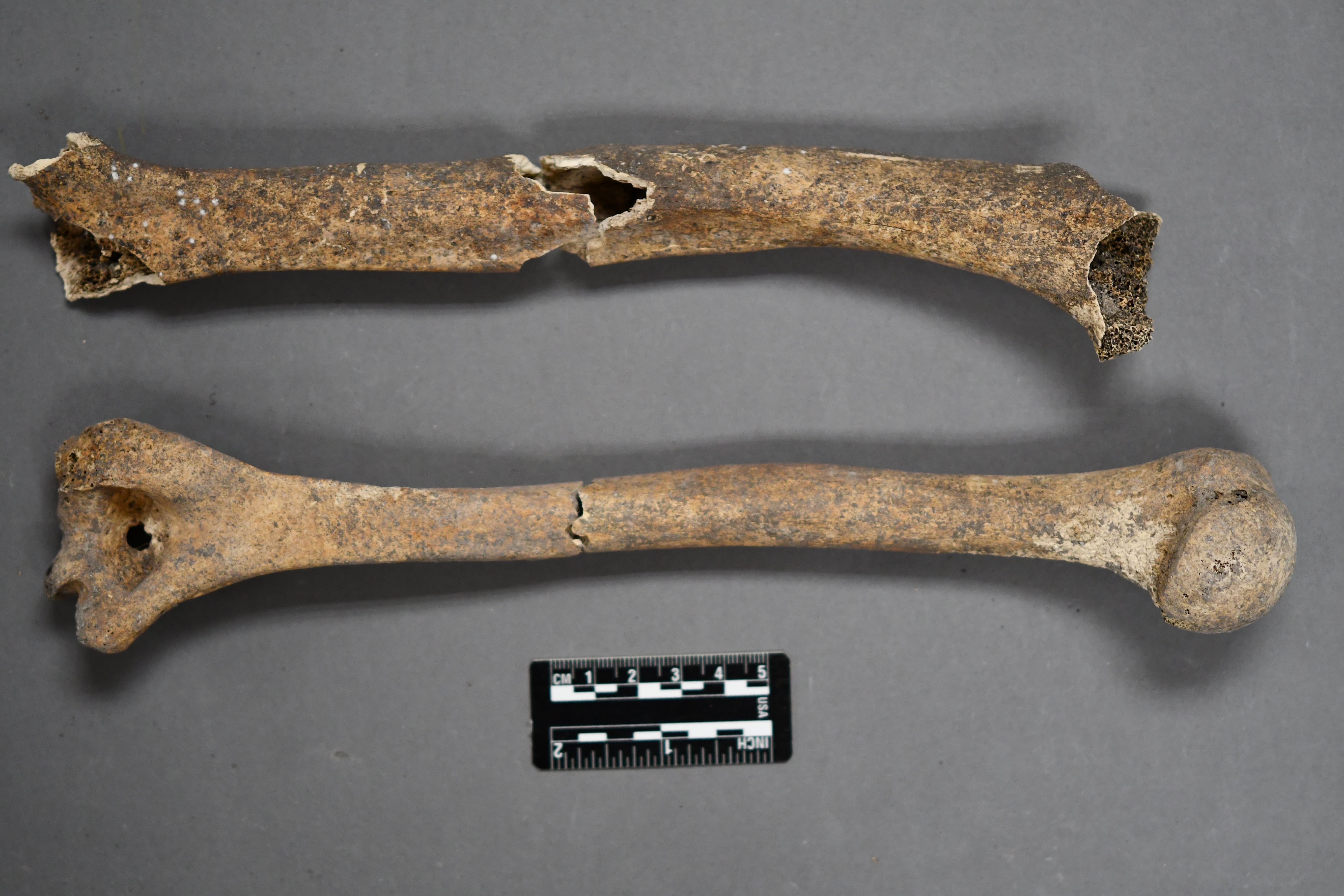
Fig 7. Humerus Comparison 1
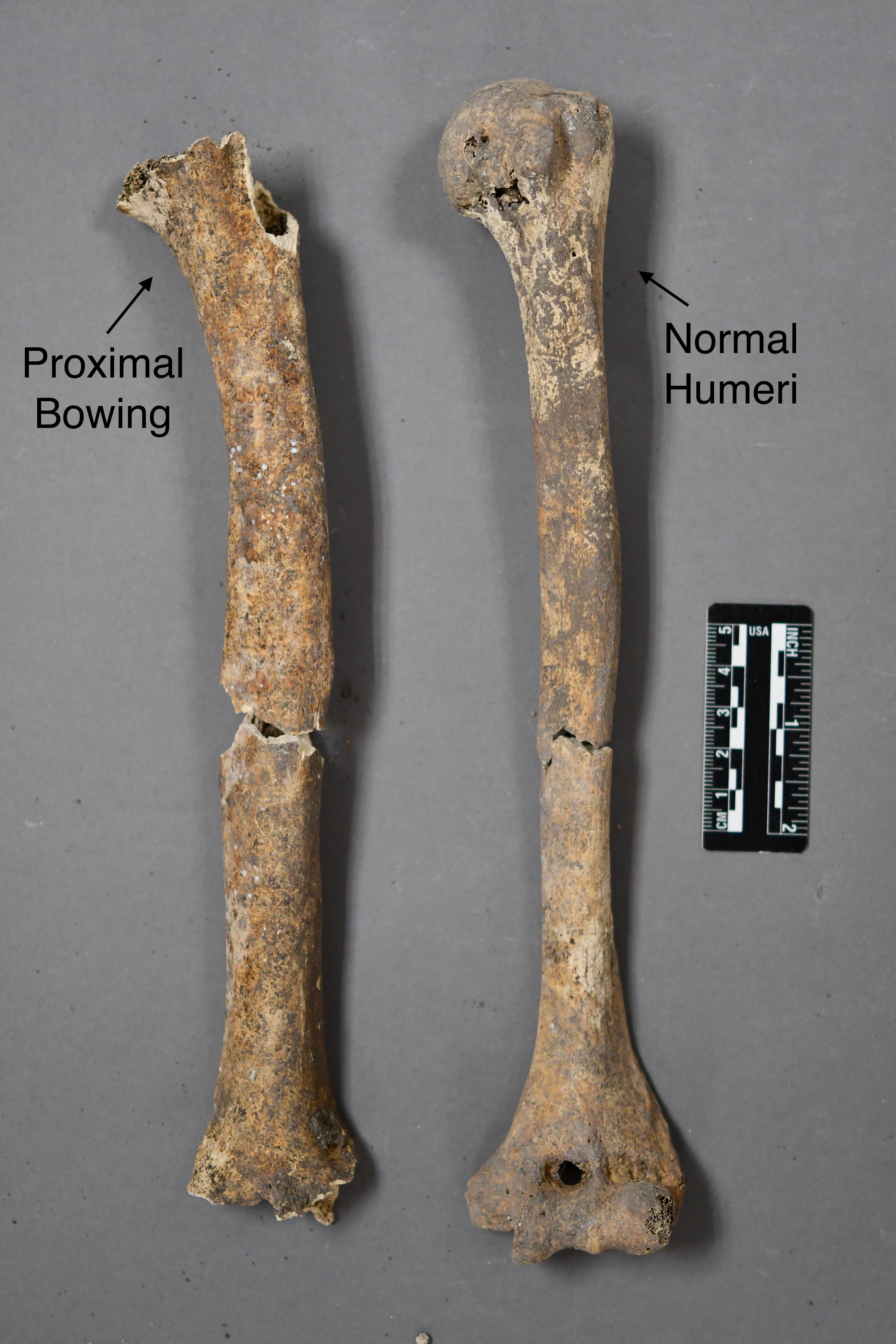
Fig 7. Humerus Comparison 2
.jpg)
Fig 8. Adult Left Radii Comparison
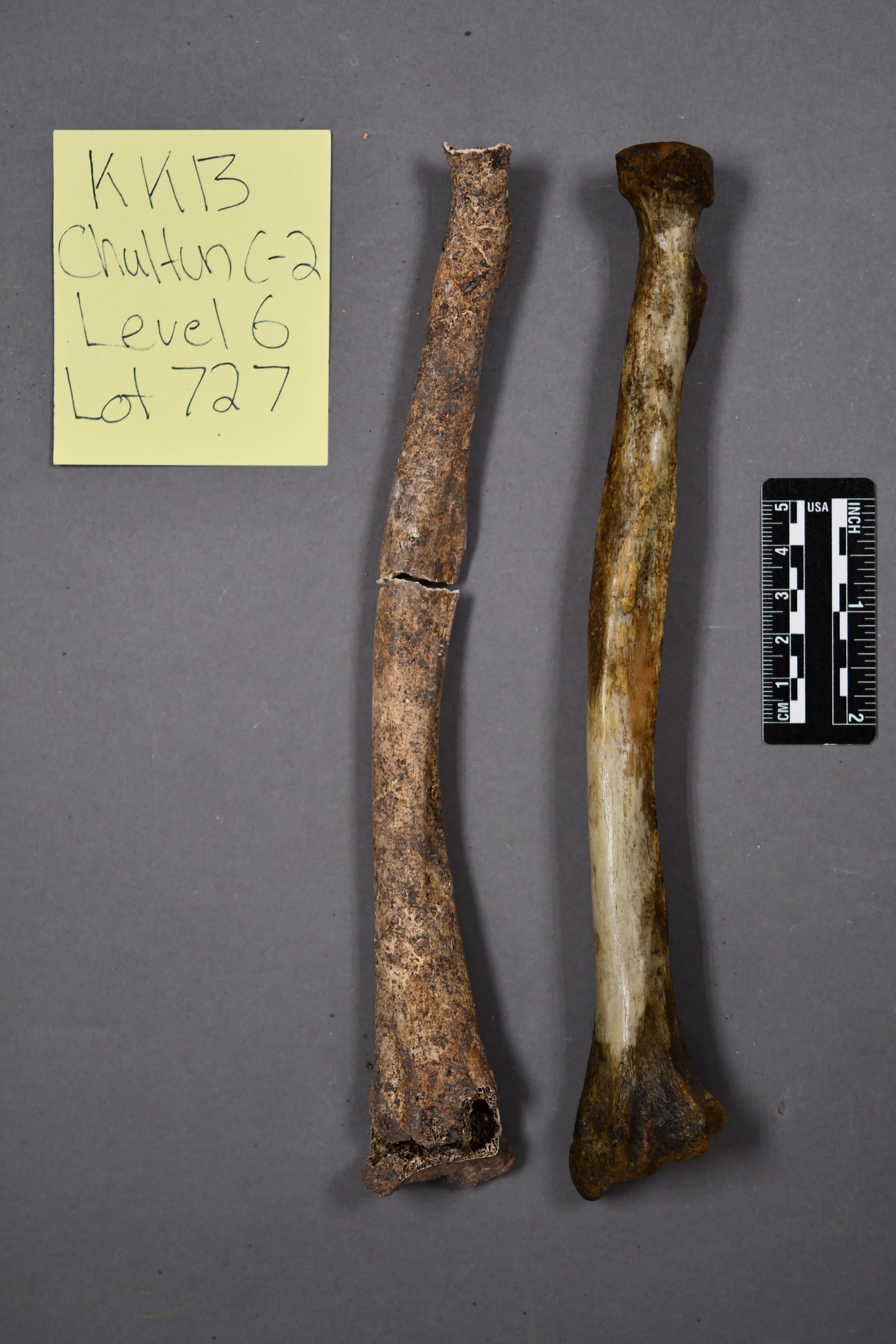
Fig 8. Adult Left Radii Comparison 2
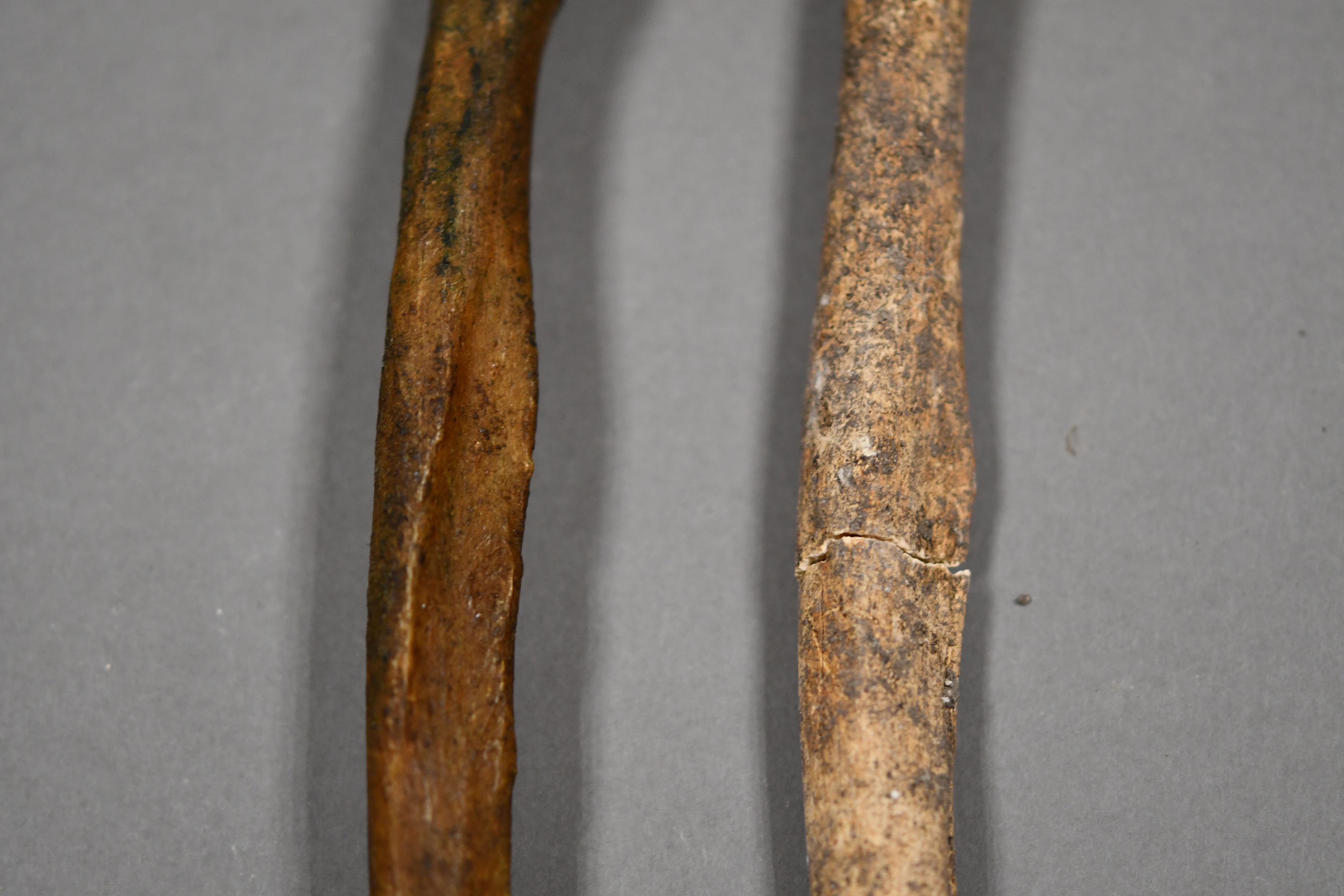
Fig 8. Left Radii Comparison Mid
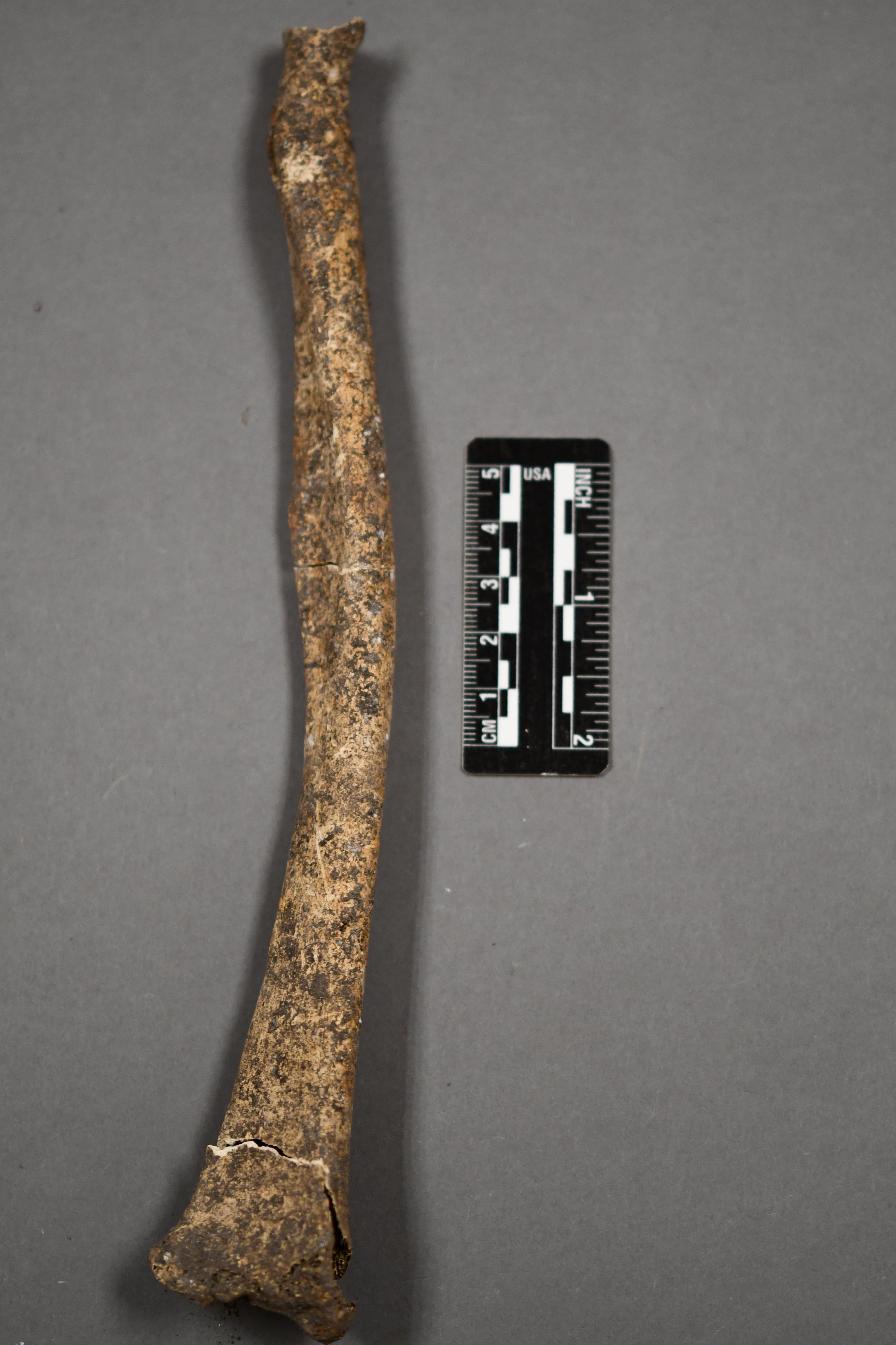
Fig 8. Left Radii
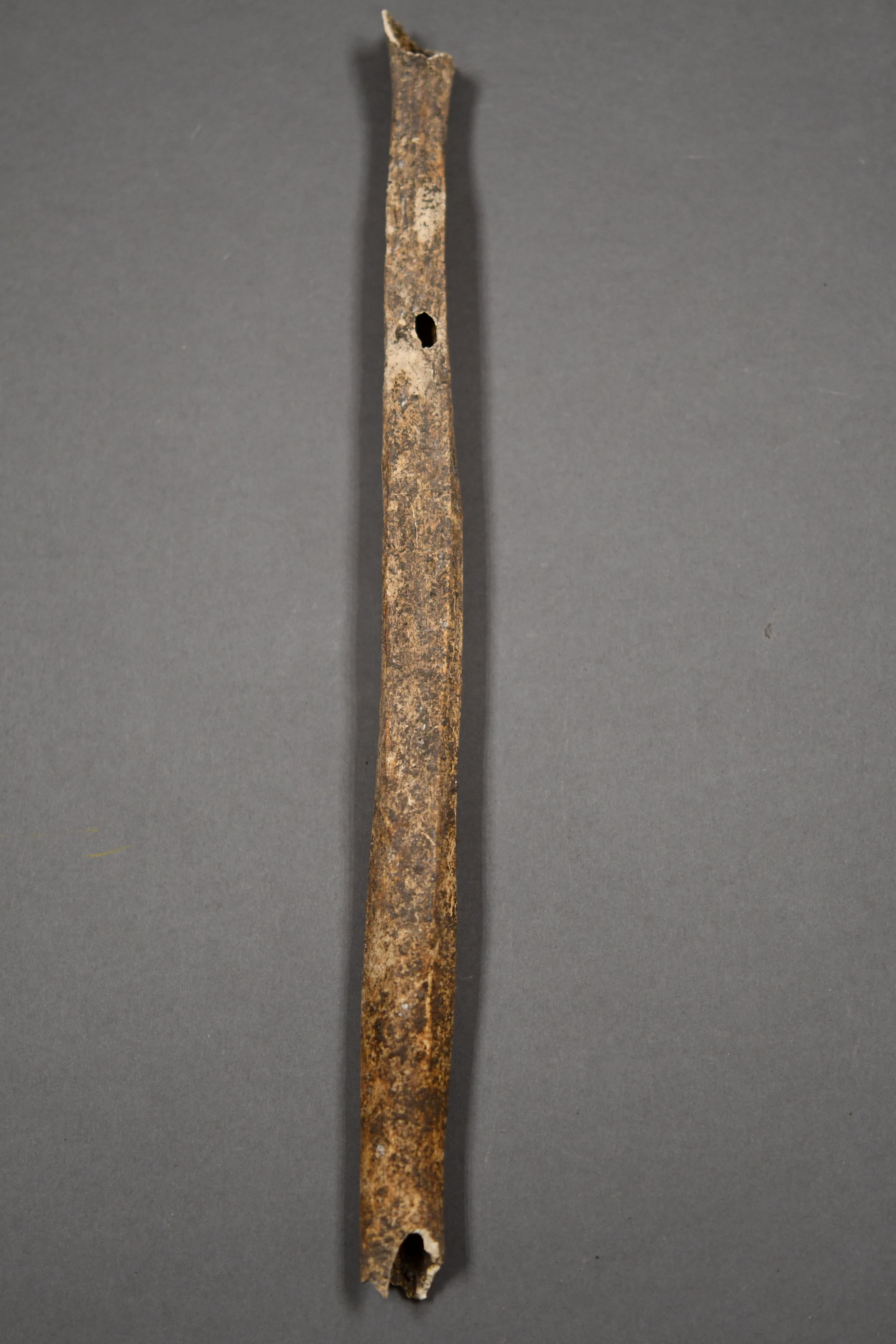
Fig 9. Adult Left Fibula
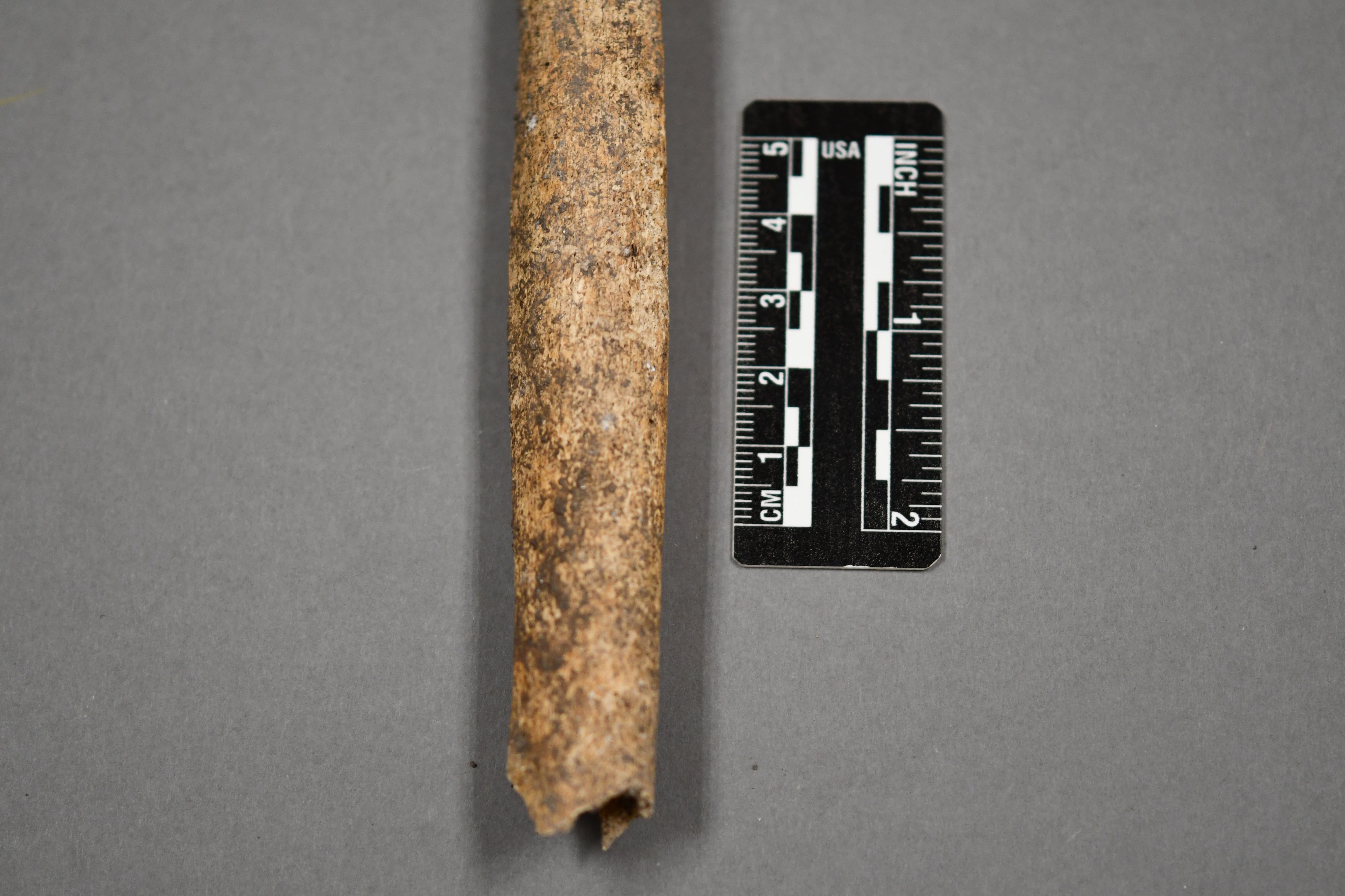
Fig 9. Adult Left Fibula, Distal
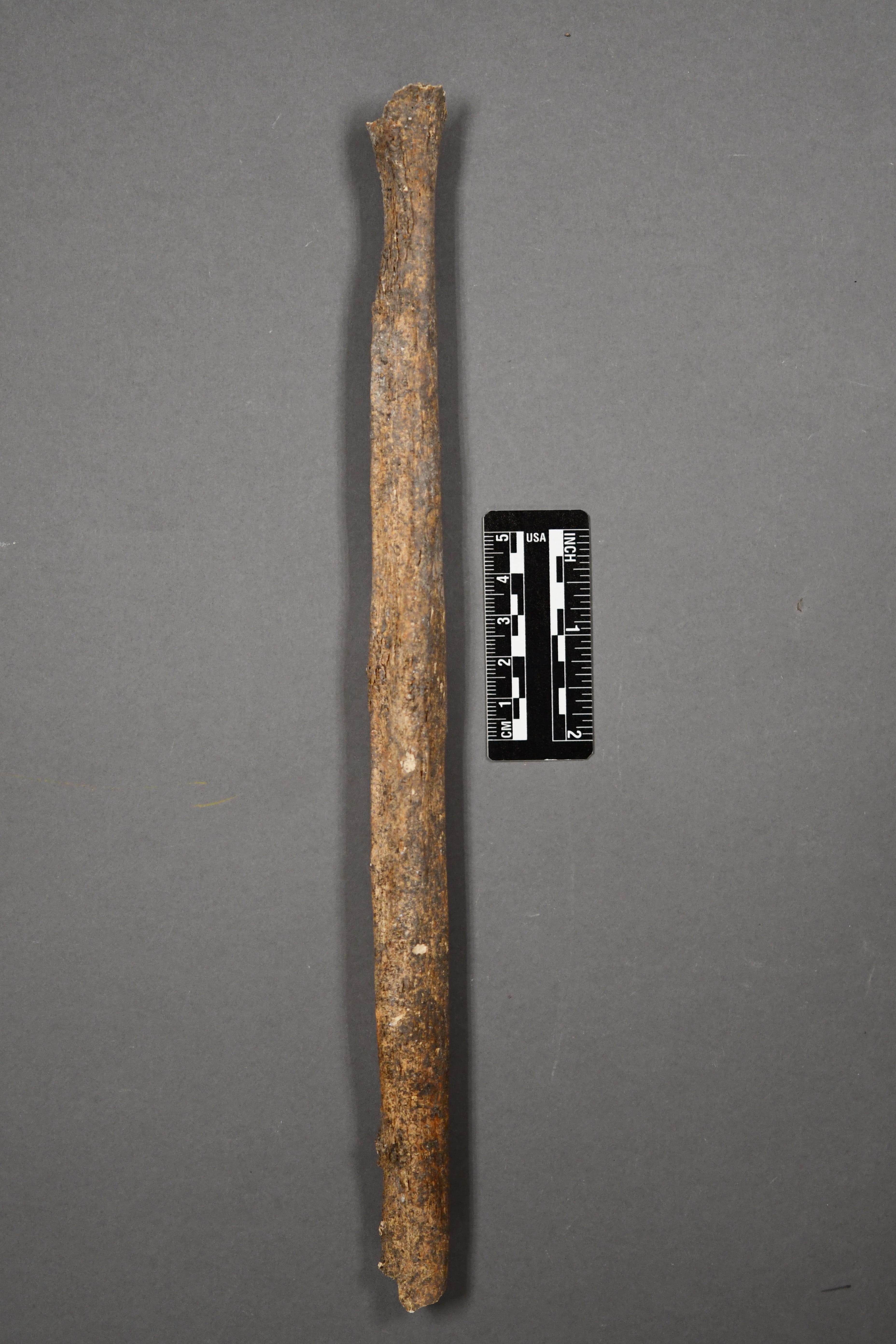
Fig 9. Adult Right Fibula
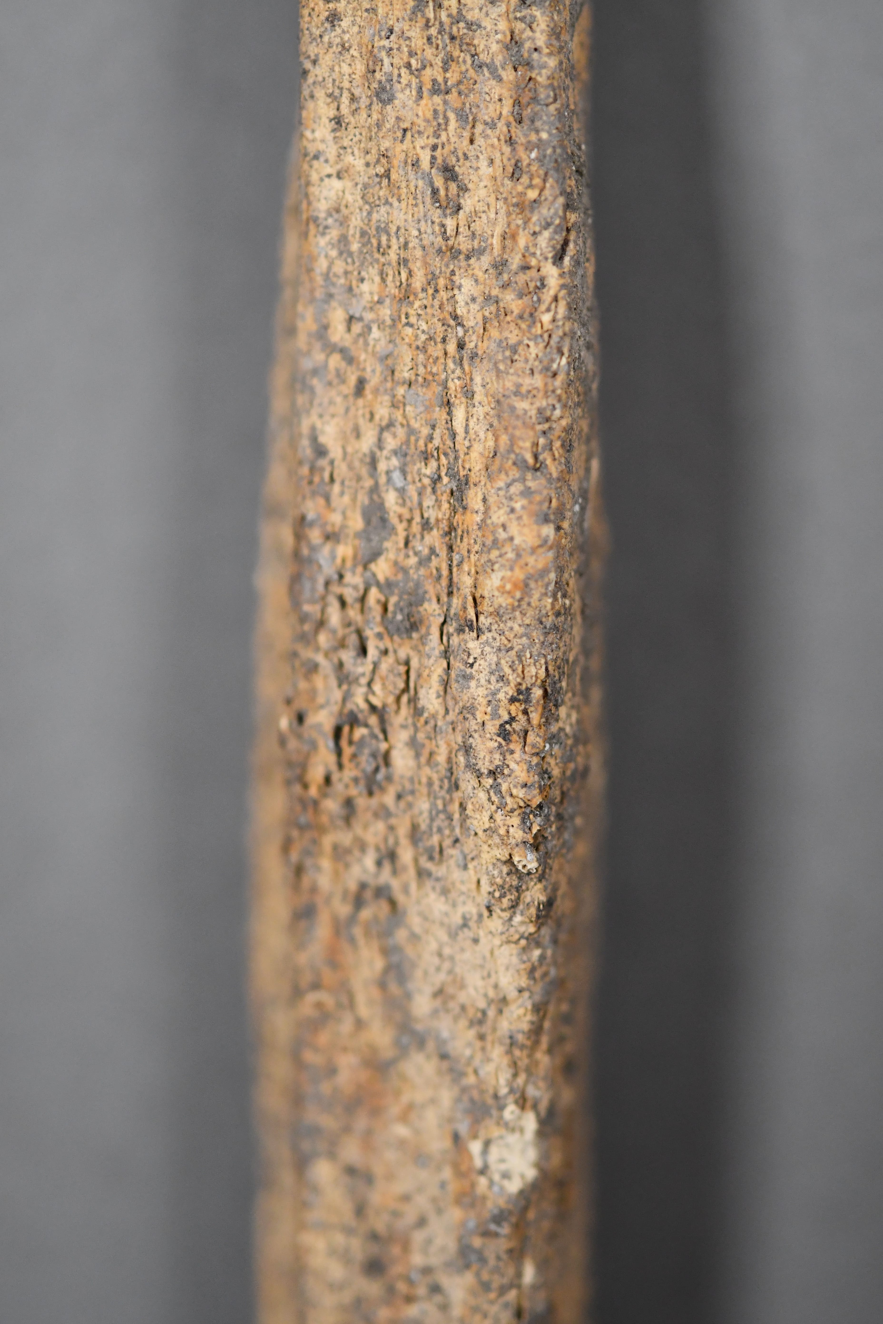
Fig 9. Adult Right Fibula Midshaft
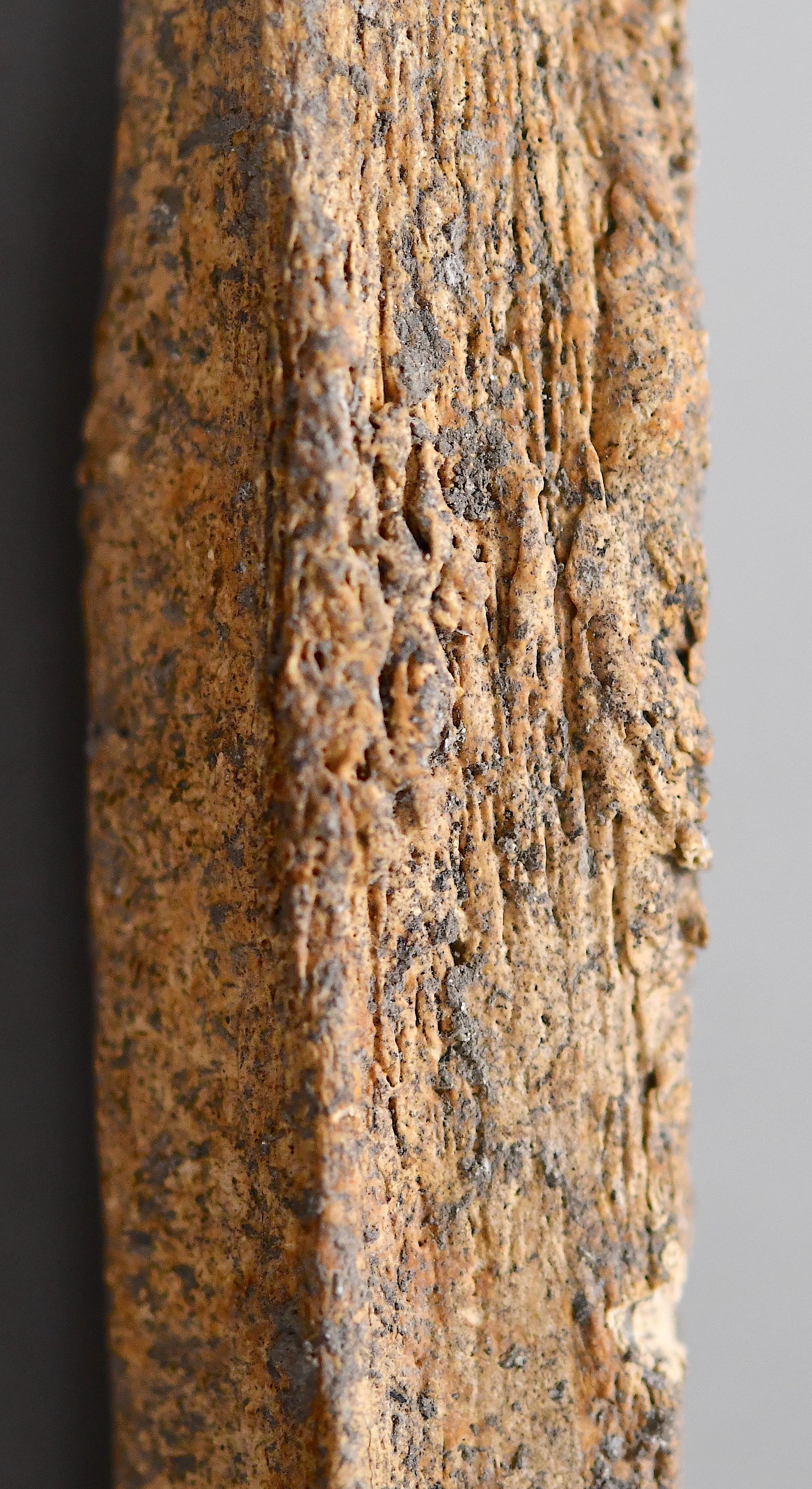
Fig 9. Adult Right Fibula Midshaft Closeup
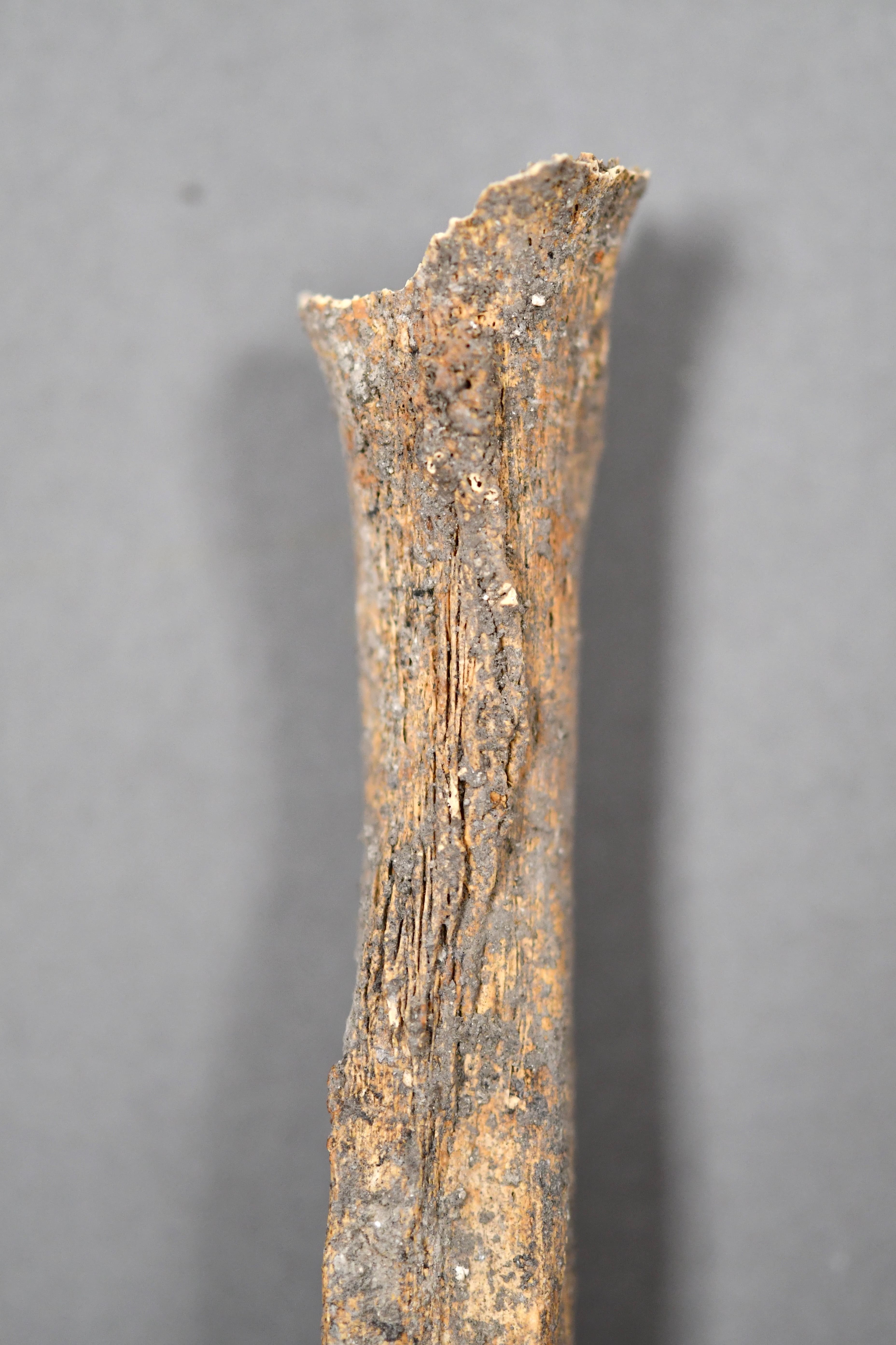
Fig 9. Adult Right Fibula Proximal